28 Which of the Following Regulates Myocyte Contraction
Notably during contraction these two heads seem to work via a hand-over-hand action such that the myosin dimer never fully lets go of the thin filament during the activation period. HuR Regulates GPCR-Mediated Intracellular Calcium Increase Through RGS.

Labeled Human Body Koibana Info Sistema Muscular Humano Anatomia E Fisiologia Humana Sistema Muscular
Cells were paced at a 500 ms cycle length for AdGFP n 26 or AdKChIP KD n 28 treated myocytes.

. A A vasoactive peptide that results in natriuretic diurises and vasodilation B Bodys endogenous defence against hypervolemia and hypertension C An N-terminal pro-Btype naturetic peptide D A group of proteins found in skeletal cardiac muscle fibres that regulate muscle contraction 39. 2014 Jun 1306 11H1525-39. Accepted in final form 21 March 2014 McCain ML Yuan.
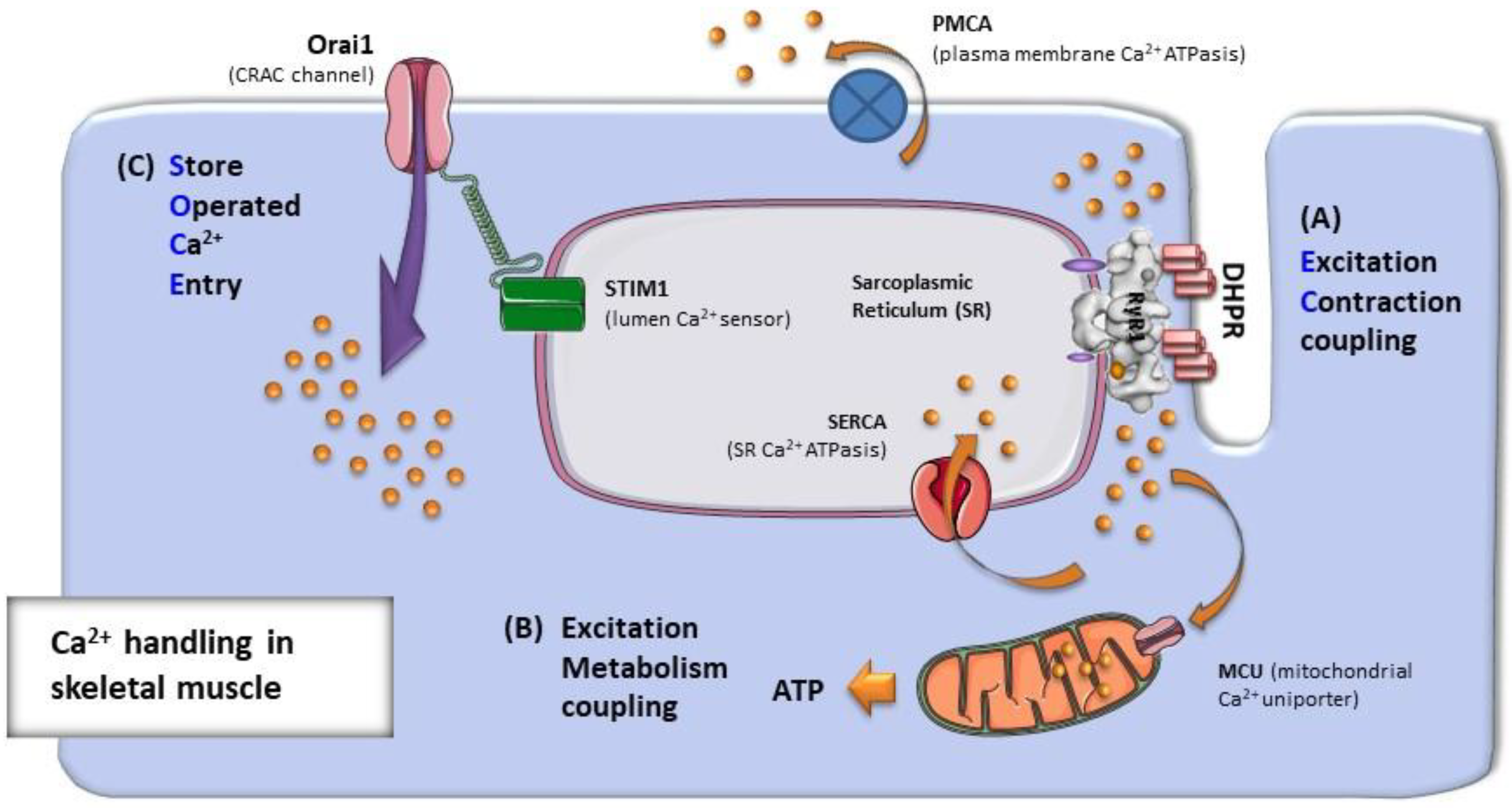
In all three muscle types the most important cellular event in relaxation is. Sarcoplasmic reticulum as the source of calcium for excitation-contraction coupling. Estrogen also regulates nNOS expression which in turn regulates cardiomyocyte contraction 101102103 104.
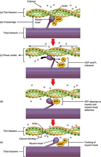
The free myosin head splits ATP. The reduction in the amount of free intracellular calcium. Both smooth and cardiac muscle cells are under hormonal control.
Pressure overload leads to cardiac hypertrophy which is often followed by heart failure. Ca 2 is the key signaling molecule for SMC contraction. Most often observed in smaller resistance arteries this basal myogenic.
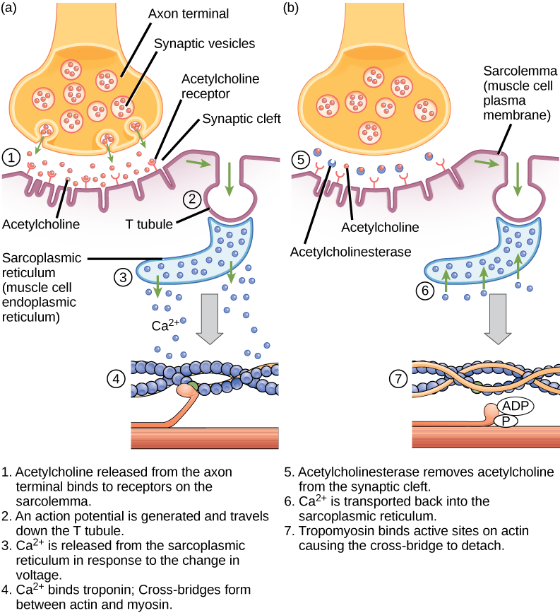
Calcium ion binds to troponin. How the electrical signal gets to each cell C. Ment regulate myocyte function.
Regulation of excitation-contraction coupling in mouse cardiac myocytes. Which of the following is not a. The myogenic mechanism is how arteries and arterioles react to an increase or decrease of blood pressure to keep the blood flow constant within the blood vessel.
Third we show that TrpC3 is a common element in the signal transduction cascades activated by. To evaluate excitation-contraction signaling pathways primary VSMCs from CTR and HuR SMKO mice were treated with 1 mmolL PE and cytosolic Ca 2 content was measured by fluorimetry. McCain Hongyan Yuan Francesco S.
Cardiac muscle contraction and heartbeat is regulated by a process known as excitationcontraction coupling ECC. A Representative tracing of the change in distance between two consecutive sarcomeres during contraction. Cyclic GMP signaling and regulation of SERCA activity during cardiac myocyte contraction Cell Calcium.
Up to 10 cash back Stained myocytes were stimulated with 20 volts at 1 Hz MyoPacer. In addition to eNOS nNOS is cardioprotective largely. Both skeletal and cardiac muscle contraction are under autonomic nervous control.
IonOptix Westwood MA USA and Ca 2 transients were recorded ratiometrically all as previously described 15 17. KChIP2 knock down reduces myocyte contractility. Up to 256 cash back Describe the following for skeletal smooth and cardiac myocytes.
Myocyte aspect ratio decreases during the compensatory stages of concentric hypertrophy 23 24 suggesting that cell shape remodeling might be an adaptive response to the microenvironment becoming stiffer. To test how myocyte shape and matrix. Myocyte contraction was recorded independently by edge detection of unloaded cardiomyocytes at 250 Hz.
To test how myocyte shape and matrix elasticity. The addition of PE evoked a transient. Myocyte aspect ratio decreases during the compensatory stages of concentric hypertrophy 23 24 suggesting that cell shape remodeling might be an adaptive response to the microenvironment becoming stiffer.
During systole depolarization of the plasma membrane opens LTCCs causing an influx of a. Tracings were normalized to control cells. Am J Physiol Heart Circ Physiol.
In vitro work has also re-vealed that cytoskeletal architecture 6 22 and contractile force generation 36 are dependent on myocyte shape another important regulator of myocyte phenotype 42 48. Contraction was induced by stimulation as. KChIP2 knock down reduces myocyte contractility.
Matrix elasticity regulates the optimal cardiac myocyte shape for contractility. As TrpC3 is localized in a subcellular domain that is different from the localization of Ca V 12 this provides strong evidence for the idea that Ca 2 influx through different ion channels differentially regulates contraction and hypertrophy in ventricular myocytes. Which of the following best describes Troponin.
The sodiumcalcium exchanger sodium pump and mechanism of action of digitalis. The myosin head pivots toward the center of the sarcomere. A kinase that phosphorylates the thick filament.
We tested the hypothesis that depressed contractility in thi. Matrix elasticity regulates the optimal cardiac myocyte shape for contractility Matrix elasticity regulates the optimal cardiac myocyte shape for contractility Megan L. Integrative analysis with mathematical modelling.
The optimal myocyte shape for contractility is regulated by the elasticity of the microenvironment. Calcium ion is released from the sarcoplasmic reticulum. Campbell Kevin Kit Parker 2014-06-01 000000 Submitted 14 October 2013.
Here we asked how both the elastic modulus of the extracellular matrix and myocyte shape regulate contractility. Regulation of cardiac contraction and relaxation by calcium. A short summary of this paper.
What is the correct sequence of these events. 10 There are two myosin isoforms in cardiac myocytes alpha and beta which have similar molecular weight but exhibit substantially different cross-bridge cycle and ATPase rates. The optimal myocyte shape for contractility is regulated by the elasticity of the microenvironment.
Myosin cross-bridges bind to the actin. Full PDF Package Download Full PDF Package. Which of the following is NOT a correct comparison of cardiac myocytes to other muscle cell types.
In cardiac and skeletal muscle cells actin and myosin are organized into sarcomeres. Myogenic response refers to a contraction initiated by the myocyte itself instead of an outside occurrence or stimulus such as nerve innervation. A Representative tracing of the change in distance between two consecutive sarcomeres during contraction.
60 The following is a list of the events that occur during a muscle contraction. Troponin as the calcium receptor in excitation-contraction coupling. 51 How Ca 2 regulates cardiac myocyte contraction.
Pin On Nclex Nursing Made Easy
Muscle Contraction And Locomotion Biology 2e

Digestive System Facts Cool Kid Facts Digestive System Diagram Digestive System Digestion

The Function Of Muscle Contraction Induced Myokines The Figure Shows Download Scientific Diagram

Ca2 Activation Of Smooth Muscle Contraction Journal Of Biological Chemistry

Contraction And Relaxation In Skeletal Muscle 1 In Healthy Skeletal Download Scientific Diagram
Ijms Free Full Text Improper Remodeling Of Organelles Deputed To Ca2 Handling And Aerobic Atp Production Underlies Muscle Dysfunction In Ageing Html

Schematic Diagram Of The Regulation Of Smooth Muscle Contraction Gpcr Download Scientific Diagram
Contraction Of Cardiac Muscle Pathway Of Contraction Teachmephysiology
0 Response to "28 Which of the Following Regulates Myocyte Contraction"
Post a Comment